Description
Product Description
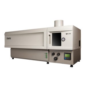
The RM5 is a compact and fully automated Raman microscope for analytical and research purposes. The truly confocal design of the RM5 is unique to the market and offers uncompromised spectral resolution, spatial resolution, and sensitivity.
Our Raman microscope builds on the expertise of robust and proven building blocks, combined with modern optical design considerations; and a focus on function, precision and speed. The result is a modern Raman microscope that stands alone in both specifications and ease of use.
- Internal Standards and Auto-Calibration – to ensure the highest quality data at all times
- 4-Position Raman Filter Turret – fully automated notch and edge filters to match the Raman range to excitation laser wavelength
- Ramacle® Software – one powerful software package for complete system control, data acquisition, analysis and ease of upgrade
- High Performance Microscope – compatible with all the latest accessories
For pricing and further information of the RM5 Raman Microscope, simply contact a member of our sales team at sales@edinst.com.
Software
RAMACLE® SOFTWARE
Ramacle is an exceptional software package written for complete instrument control and data handling on the RM5 system. Ramacle controls all RM5 functions with a straightforward design concept. It focuses on all modern Raman spectroscopy applications, while at the same time, providing a user-friendly interface with ‘ready to publish’ outputs.
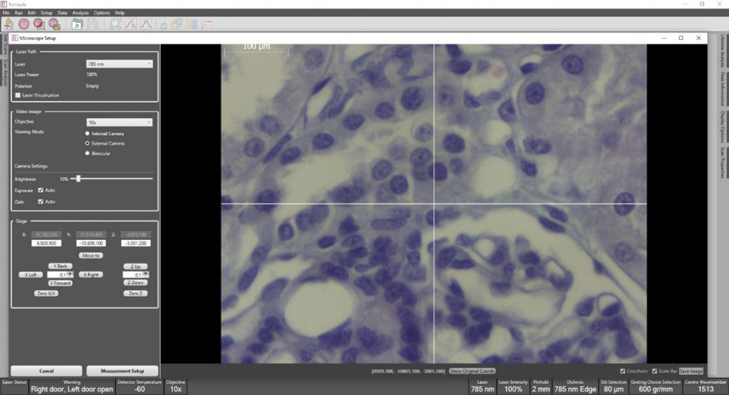
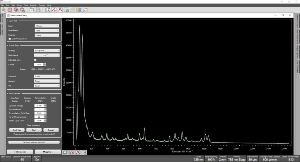
The software provides control, visualisation, data acquisition, analysis and presentation of the RM5 whether it is used for generating Raman spectra or with advanced upgrades such as Raman mapping. Ramacle easily navigates the user from microscope set up to measurement set up and offers computer-controlled measurement conditions such as laser excitation, grating, pinhole size etc.
Ramacle enables sample visualisation, live signal monitoring and parameter optimisation before every measurement. The instrument status and signal are displayed and constantly updated during measurements.
Data generated by Ramacle have a proprietary file format. This contains all measurement and instrumental properties, allowing the user to retrieve important information whenever needed and ensures data traceability. Simple input and output functions provide the required compatibility with third party data analysis or presentation packages.
KnowItAllTM Raman Identification Pro spectral library is available for material identification and advanced analysis. Data acquisition methods such as single measurements, multiple and accumulated scans, kinetic scans and generation of maps (accessory dependent) are implemented by intuitive and in user-friendly wizards.
Raman Mapping
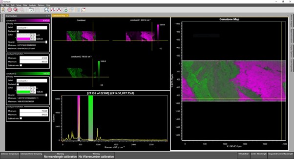
Mapping the Raman spectra within a sample area provides previously unavailable information about the chemical and physical differences across a sample. This can confirm the identity and presence of specific components and reveals their location and distribution within the sample. The figure demonstrates an XY Raman map of a gemstone containing two constituents. The Raman map is false coloured based on Raman bands specific to each constituent.
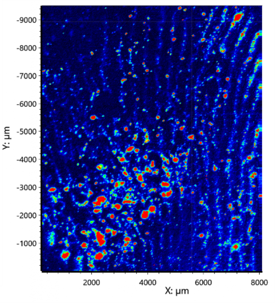
Using Ramacle the user can use our fast-mapping technique. This uses the camera and motorised stage to the fullest, greatly reducing acquisition times. The figure below shows Raman map revealing trace amounts of paracetamol (shown in red) left behind in a fingerprint on aluminium foil. Using standard mapping this map would have taken over 20 hours, using fast mapping this acquisition took under 30 minutes, without losing any spectral or spatial quality.
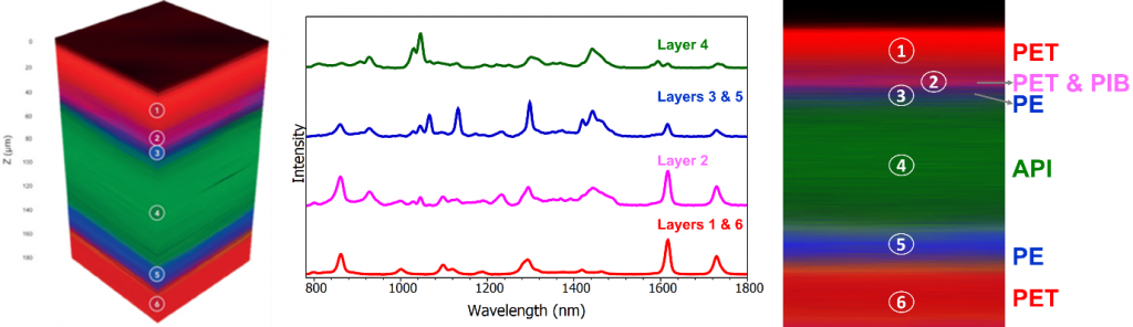
Mapping in Ramacle also allows measurements in the Z axis, thanks to the computer-controlled pinhole. 3D mapping enables the user to gain a depth profile of their sample and is extremely useful for analysing layers samples for quality control and layer thickness conformation. Below is a 3D Raman map of a transdermal patch is shown, the different layers can clearly be differentiated.
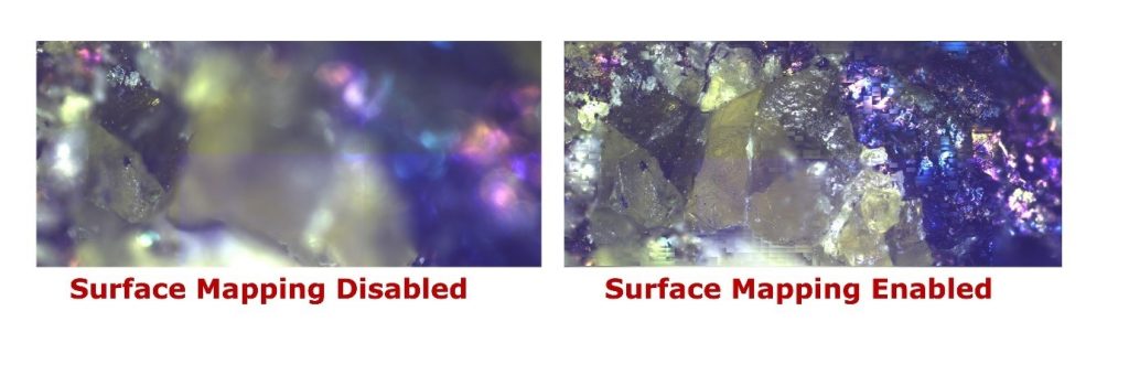
Commonly, samples are not perfectly flat, and this can cause focus issues when Raman mapping. Using the surface mapping feature in Ramacle the user can create an in-focus white light image with set Z-positions. These Z-positions will then be used for the Raman map creating a completely in focus 2D Raman map. The image below shows the difference between a sample with and without surface mapping.
Ramacle Key Features
- Selection of laser and scatter optical pathways
- Selection of excitation wavelength, gratings and exposure time
- Sample and laser focus visualisation
- Programmed attenuator and shutter
- Single, accumulated and kinetic spectral acquisitions
- Spectral correction
- Selection and scans of internal calibration standards and automated calibration correction
- Data operations such as arithmetic, scaling, normalisation and baseline subtraction
- Cosmic ray removal, cropping, smoothing
- Automated laser alignment
- ASCII / CSV data import / export function
- Paste options for presentations and publications
Features included with upgrade:
- Mapping features – map setup, fast mapping, surface mapping, 3D mapping, collection and data analysis
- Fully motorised stage – XYZ control through joystick and software
- Polariser and analyser selection and control
- Detectors selection
- Laser rejection filter selection
- External camera selection and visualisation
- Autofocus